
jumpincactus
Supporter
Premium Member
- Posts
- 11,605
- Reactions
- 35,935
- Joined
- Jul 24, 2011
- Points
- 438
Abstract
Charcoal has a long soil residence time, which has resulted in its production and use as a carbon sequestration technique (biochar). A range of biological effects can be triggered by soil biochar that can positively and negatively influence carbon storage, such as changing the decomposition rate of organic matter and altering plant biomass production. Sorption of cellular signals has been hypothesized to underlie some of these effects, but it remains unknown whether the binding of biochemical signals occurs, and if so, on time scales relevant to microbial growth and communication. We examined biochar sorption of N-3-oxo-dodecanoyl-L-homoserine lactone, an acyl-homoserine lactone (AHL) intercellular signaling molecule used by many gram-negative soil microbes to regulate gene expression. We show that wood biochars disrupt communication within a growing multicellular system that is made up of sender cells that synthesize AHL and receiver cells that express green fluorescent protein in response to an AHL signal. However, biochar inhibition of AHL-mediated cell-cell communication varied, with the biochar prepared at 700°C (surface area of 301 m2/g) inhibiting cellular communication 10-fold more than an equivalent mass of biochar prepared at 300°C (surface area of 3 m2/g). These findings provide the first direct evidence that biochars elicit a range of effects on gene expression dependent on intercellular signaling, implicating the method of biochar preparation as a parameter that could be tuned to regulate microbial-dependent soil processes, like nitrogen fixation and pest attack of root crops.
Keywords: biochar, carbon sequestration, charcoal, microbial communication
Go to:
Introduction
Charcoal is a ubiquitous component of the Earth system, present not only in the soils of fire-affected ecosystems, but also in marine sediments and terrestrial and marine dissolved organic carbon pools.1 It also forms the basis for a form of carbon sequestration called soil biochar amendment,2 where charcoal is intentionally added to soil with the aim of increasing the size of the stable organic carbon pool while potentially delivering other agronomic benefits such as increased crop productivity,1,3–5 decreased soil tensile strength,2,6 altered soil water properties,7–10 improved plant pest resistance,11 and decreased nitrogen loss from soils.12 The carbon sequestration potential of biochar is based the assumption that a large fraction of this material is an inert component of the soil organic matter pool with a very long soil residence time.13 Large-scale data on the biogeochemical cycling of charcoal back up the idea that it decomposes slowly in the Earth system,14,15 and laboratory and ecosystem studies are narrowing down the factors that control charcoal soil residence time.16–20
As commercial applications of biochar have begun, results have appeared which challenge the inert nature of this material and suggest more complex carbon cycle roles. For example, biochar addition to soil has been shown to promote the loss of non-charcoal organic matter (priming) in some studies,21 but not in others.22 This priming of soil decomposition causes an enhanced flux of CO2 into the atmosphere from soils, potentially decreasing the carbon sequestration benefits of biochar, as well as decreasing ecosystem services provided by soil organic matter. Other evidence has appeared suggesting that biochar can enhance retention of nitrogen molecules that contribute to soil fertility,23,24 stimulate colonization of roots by mycorrhizal fungi,25,26 change soil microbial composition,27 and confer plant resistance to microbial pathogens.28 These biological effects have been proposed to arise in part because biochars sorb diffusible small molecules that soil organisms use for intercellular communication and coordinated decision-making.25,29Biochars are known to bind diverse organic molecules,30 many of which are nonpolar like the molecules used for intercellular communication. Numerous studies have analyzed the kinetics of biochar sorption to organics on time scales (days to months) relevant to the mobilization of pollutants.29,31–33 However, it remains unclear if communication can be altered by the presence of biochars on the time scales of microbial signaling and gene expression (minutes to hours).
It is challenging to directly demonstrate biochar-driven mechanisms for observed biological effects within the environment because of the complexity and diversity of conversations occurring among microbes and plants. Bacteria communicate with one another using a variety of biochemicals,34 which are distinct from the molecules used by fungi for intraspecies communication.35 Plants also synthesize flavinoids that regulate microbial behaviors, such as the establishment of root nodules,36 and microbes synthesize nodulation signals37 and plant hormones38 that influence plant development and nutrient uptake. The diversity of signals present in the environment creates challenges in attributing causality to any particular intercellular conservation. The high light absorptivity of biochar and soils adds to this challenge. While there exist microbial biosensors capable of synthesizing reporters that can be imaged when they encounter biological signals within the rhizosphere,39,40fluorescent reporters tend to absorb and emit light in a visible range that is unsuitable for imaging cells within biochar-amended materials, which have high light absorption.
To better understand the effects of biochars on cellular communication, we investigated how biochar materials created under different pyrolysis temperatures (300, 350, 400, 450, 550, 600, and 700°C) influence signal detection within a synthetic microbial system.41 Because the relevant question is the detectability of molecular signals by microbes (as opposed to the chemically-extractable fraction of signal present in the soil), we used a microbial sensor to determine when signaling molecules fell below a level detectable to bacteria in the presence of different biochars. We focused our attention on N-3-oxo-dodecanoyl-L-homoserine lactone, a member of the acyl-homoserine lactone (AHL) signaling molecule family that are used for intraspecies communication and quorum sensing by many gram-negative bacteria, including nitrogen fixing plant symbionts and pathogens that cause soft rot in plants.42,43
Go to:
Materials and Methods
Materials
Escherichia coli XL1-Blue were from Stratagene, and E. coli BLIM cells were kindly provided by K.S. Matthews.44 The acylhomoserine lactone (AHL) used, N-3-oxo-dodecanoyl-L-homoserine lactone, was from Cayman Chemical, bacterial growth media components were from BD Biosciences, and all other reagents were from Sigma-Aldrich and VWR.
Biochar synthesis
Slow pyrolysis of Prosopis glandulosa (mesquite) wood to generate biochar was performed using a fixed bed reactor as described previously.7 In brief, mesquite feedstocks ground to 20 mesh (<0.853 mm) were placed in a stainless steel crucible, which was then plugged with ceramic wool, capped with a ceramic bowl, and buried in fine-grained quartz sand inside a larger, open-top stainless steel crucible. This reactor system was heated in a muffle furnace at 5°C min−1 to the desired reaction temperatures (300, 350, 400, 450, 550, 600, and 700°C) and held at each temperature for 4 hours. The products were manually mixed after cooling to increase homogeneity.
Surface area measurements
We measured the biochar surface area using a Quantachrome Autosorb-3b Surface Analyzer. Prior to analysis, samples were placed in ashed (550°C for 4 hours) glass cells and vacuum dried overnight at 200°C. Nitrogen adsorption/desorption isotherms were obtained at 77 K by a 26-point analysis for relative pressures P/P0from 1.21 × 10⁻⁴ to 0.99, where P is the adsorption equilibrium pressure and P0 is the vapor pressure of bulk liquid N2 at the experimental temperature. Specific surface area was calculated using Brunauer-Emmett-Teller (BET) theory.45
Receiver plasmids
Receiver plasmids contained the synthetic PlasR promoter fused to a strong RBS (BBa_B0034) and GFP gene (BBa_E0040) from the IGEM registry. These plasmids additionally contained a pMB1 origin from pET28a and either a chloramphenicol (CmR) or a kanamycin (KanR) selectable marker.
Sender plasmid
The sender plasmid contained the PA1lacO-1 promoter46 fused to a strong RBS and a gene fusion that was made up of the LasI gene (BBa_C0078) fused to the mCherry gene through a (GGGGS)3 peptide linker. This plasmid additionally contained a pMB1 origin, a CmR marker, and the LacI gene from pET28a. In this sender plasmid, the N-3-oxo-dodecanoyl-L-homoserine lactone synthase gene, LasI, is expressed as a fusion to mCherry through a (GGGGS)3 linker using a PA1lacO-1promoter, which is constitutively active in cells that lack the repressor LacI, such asE. coli BLIM cells.44 The function of this fusion protein is indistinguishable from LasI.
Effect of AHL on GFP expression in liquid culture
E. coli XL1 Blue harboring the receiver plasmid (CmR) were grown to stationary phase in LB medium containing 34 Mg/mL chloramphenicol, diluted to an A600 = 0.05 in 50% LB medium that contained varying concentrations of AHL (0, 5, 25, 50, 125, 250, 500, 1000, and 2000 nM), and grown to stationary phase at 30°C. GFP levels were determined by measuring whole cell fluorescence emission (509 nm) upon excitation of 488 nm using a Tecan M1000 plate reader. Fluorescence was normalized to culture absorbance at 600 nm to account for variability in growth.
Effect of biochar on AHL availability in water
Varying concentrations of each biochar (1, 5, 10, 25, and 50 mg/mL) prepared at different temperatures (300, 350, 400, 450, 550, 600, and 700°C) were incubated with 1 MM AHL within water for 6, 60, or 1440 minutes. After incubation, biochar was removed through centrifugation (14,000 g for 1 min), and 100 ML of the soluble fraction was mixed with an equal volume of LB medium containing E. colitransformed with the CmR receiver plasmid at an OD600 = 0.05 and 50 Mg/mL chloramphenicol. This mixture was grown for 18 hours within 96-well microtiter plates incubated at 30°C and shaken at 250 rpm. Green cellular fluorescence (λex = 488; λem = 509) and absorbance (600 nm) were measured using 200 ML of each culture after growing cells in a shaking incubator (30°C; 250 rpm) for 18 hours. To account for variation in cell density, fluorescence to absorbance ratio was calculated for each well, and the calculated values were normalized to those observed with cells that were grown in the absence of AHL and in the presence of AHL that was not incubated with a biochar.
Effect of colony separation on intercellular signaling
E. coli XL1 Blue harboring the KanR receiver plasmid and E. coli BLIM transformed with the CmR sender plasmid were grown to stationary phase in LB containing 50 Mg/mL kanamycin and 50 Mg/mL chloramphenicol, respectively. Bacteria (1 mL) were harvested by centrifugation, washed with 1 mL of 25% glycerol, resuspended in 1 mL of 25% glycerol, and diluted 10-fold in 25% glycerol. Resuspended receiver (8 ML) and sender cells (3 ML) were spotted at different distances (1, 10, 20, and 30 mm) from one another on 1.5% agar plates containing M9 minimal medium and twenty amino acids. A smaller volume of the sender strain was spotted to avoid growth of these cells over the biochar agar slabs during the overnight incubation. Spotting was performed in triplicate at each separation distance. After spotting, plates were incubated overnight at 37°C and then photographed with a Canon Digital Rebel mounted on a Leica MZFLIII microscope at 0.4x magnification with white light (5 ms exposure), a Chroma 41012 GFP long pass filter set (200 and 500 ms exposures), and a Chroma 11002 Green filter (200 and 500 ms exposures). The image analysis software ImageJ was used to quantify the fluorescence from receiver colonies on each agar plate.47
Effect of biochar on cell-cell communication on solid medium
Agar plates for assessing cellular communication were prepared by placing a mold (sterilized through immersion in ethanol) within sterile polystyrene plates (60×15 mm) and pouring 8 mL of 1.5 agar containing M9 medium and twenty amino acids. Upon cooling the mold was removed to leave a pair of empty adjacent wells. The left well (control) on each plate was filled with 1 mL liquid agar (1.5%) dissolved in water (65°C). The right well (experimental) was then filled with 1 mL liquid agar containing or lacking 10 mg of 300°C or 700°C biochar. E. coli XL1 Blue harboring the KanR receiver plasmid and E. coli BLIM transformed with the CmR sender plasmid were grown to stationary phase in LB containing 50 Mg/mL kanamycin and 50 Mg/mL chloramphenicol. Bacteria were harvested by centrifugation, washed with 25% glycerol, resuspended in 25% glycerol, and diluted 10-fold into 25% glycerol. The 10-fold dilution of sender cells (3 ML) was spotted between the two agar slabs on plates, whereas the 10-fold dilution of receiver cells (8 ML) was spotted on the outside of each agar slab. After spotting, plates were incubated overnight at 37°C and then photographed with a Canon Digital Rebel on a Leica MZFLIII microscope at 0.4× magnification with white light (5 ms exposure), a Chroma 41012 GFP long pass filter set (200 and 500 ms exposures), and a Chroma 11002 Green filter (200 and 500 ms exposures). ImageJ was used to quantify the fluorescence from the pair of receiver colonies on each agar plate. The ratio of the signals from the pair of receiver colonies on each plate was calculated (receiver adjacent to biochar/receiver adjacent to empty agar) to determine the level of signal inhibition elicited by each biochar. All measurements were performed in triplicate, and values reported represent the average of three independent experiments ±1σ.
Calculating the effect of biochar on AHL diffusion
To estimate the effect of biochar on AHL diffusion within agar plates, the effective diffusion coefficient De of AHL was calculated as (1 − ε)D0/[1 − 0.5 ln(1 − ε)], where D0 is the actual diffusion coefficient in biochar-free agar medium and is the volume fraction occupied by biochar particles in the agar-BC cube.48 This analysis revealed that the low volume fraction used in our experiments (less than 2% v/v biochar) decreases the AHL diffusion constant by less than 3%. This implicates sorption as the major mechanism responsible for biochar effects on sender-receiver cell-cell signaling.
Go to:
Results and Discussion
Autoinducer sorption to biochars
To assay whether biochars influence the availability of AHLs, we constructed a bacterial biosensor39 that expresses a green fluorescent protein (GFP) reporter when bacterial cells encounter the AHL N-3-oxo-dodecanoyl-L-homoserine lactone in their local environment. This was accomplished by transforming E. coli with a plasmid that: i) constitutively expresses the protein LasR, a Pseudomonas aeruginosa AHL-dependent protein that activates transcription from the promoterPlasR, and ii) expresses GFP under control of the LasR-dependent promoter PlasR. Cells containing this receiver plasmid displayed increased GFP production when they were grown in medium containing AHL (Fig. S1), because LasR requires bound AHL to activate transcription of genes whose expression is regulated by the LasR promoter.49 We assayed AHL signaling in E. coli because this organism does not use AHL to communicate, lacks the complex responses of soil bacteria that are sensitive to one or more AHL, and does not produce lactonase enzymes that hydrolyze AHL. With this bacterial biosensor, only two parameters determine AHL levels in the medium: the amount of AHL added to the medium and fraction of the AHL that sorbs to biochar.
We produced a suite of biochars by pyrolyzing Prosopis glandulosa (mesquite) wood chips over a range of temperatures (300, 350, 400, 450, 550, 600, and 700°C). N2 sorption analysis of these biochars revealed surface areas that spanned more than two orders of magnitude (Fig. S2). Varying amounts of each biochar (1 to 50 mg/mL) were incubated with a concentration of AHL (1 MM) sufficient to activate E. coli to synthesize GFP (Fig. S1). This level of AHL is within a range that is thought to be biologically relevant for Pseudomonas aeruginosa.50 Initial incubations were performed on the timescale of E. coli reproduction. After 60 minutes, the biochar was separated from unbound AHL by centrifugation, the soluble fraction was mixed with a low density culture of E. coli, and cells were grown to stationary phase to allow for GFP expression. Whole cell GFP fluorescence measurements revealed that incubation of AHL with the highest concentrations of each biochar decreased AHL-dependent cellular fluorescence in all cases (Fig. 1). The concentration of biochar required to suppress cellular fluorescence varied with biochar pyrolysis temperature and surface area. The biochars with similar low surface areas (300, 350, and 400°C) required the highest concentrations to suppress GFP expression half maximally (10–50 mg/mL), the biochars with the highest surface areas (550, 600, and 700°C) required the lowest concentrations to suppress GFP expression half maximally (1–2 mg/mL), and the biochar with an intermediate surface area (450°C) required an intermediate level of biochar (5 mg/mL) to suppress GFP expression half maximally. We also found that the correlation between biochar surface area and the concentration required for half maximal inhibition of GFP fluorescence could be described by a simple exponential model, R2 = 0.99 (Fig. 2). This trend implicates the AHL binding capacity of the biochar as dependent on surface area and pyrolysis temperature under the conditions used to produce our biochars. Cells incubated with biochar-treated AHL all grew to a similar maximal density (Fig. S3), indicating that biochar-treated water did not alter cell growth.
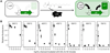
Figure 1
Sorption of AHL by biochars pyrolyzed over a range of temperatures. (A) E. coli transformed with the receiver plasmid synthesize GFP when grown in an environment containing the AHL N-3-oxo-dodecanoyl-L-homoserine lactone. The transcriptional activator ...
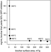
Figure 2
Relationship between biochar surface area, production temperature, and inhibition of GFP reporter expression. The concentration of biochar required to decrease AHL-induced GFP fluorescence by 50% within whole cells was estimated from the data presented ...
We also sought to establish whether the biochar sorption of AHL had reached equilibrium with our low surface area biochars and whether biochar sorption occurred on an even faster time scale with our high surface area biochars, e.g., faster than the doubling time of E. coli in rich growth medium. If some biochars sorb AHL on the timescale of very rapidly-growing E. coli under optimal growth conditions in a lab, they are likely to sorb AHL under the lower nutrient, slower growth conditions experienced by soil microbes in the environment. To address these questions, we varied the incubation time of AHL and biochar prior to mixing the soluble fraction with E. coli. Incubation of low temperature biochars (300 to 400°C) with AHL for 24 hours showed a greater inhibition of GFP expression than 1 hour incubation (Fig. S4), indicating that these reactions had not reached equilibrium after one hour. The lack of saturation suggests the ability of biochar to cause a continued suppression of microbial signaling as bacterial populations grow. In addition, AHL-induced cellular fluorescence was suppressed when the high temperature biochars (550 to 700°C) were incubated with AHL for only six minutes prior to mixing with E. coli (Fig. S5). This latter finding suggested that some biochars are capable of adsorbing AHL on the very shortest time scales of microbial reproduction and signaling within rich growth medium, a condition that likely represents the upper bound on the rate of growth and signaling within the environment.
Effect of biochars on AHL-mediated cell-cell communication
To directly assess whether biochars can disrupt communication among growing bacteria, we built a sender plasmid that programs E. coli to express P. aeruginosaLasI (Fig. 3A), the acyl-homoserinelactone synthase that continuously synthesizes the AHL N-3-oxododecanoyl-L-homoserine lactone from bacterial metabolites.51 LasI was expressed as a fusion to the red fluorescent protein mCherry to allow for visualization of expression. To make LasI production of N-3-oxo-dodecanoyl-L-homoserine lactone continuous within sender cells, we used a PA1lacO-1 promoter to constitutively express the LasI-mCherry fusion protein and E. coli BLIM cells that are devoid of the transcriptional lac repressor, which inhibits expression of genes from this promoter. To determine how receiver-sender cell-cell communication varied with distance within a synthetic multicellular system,41 E. coli cells harboring each plasmid type were spotted onto solid growth medium at different edge-to-edge distances from one another (10 to 30 mm), and cell density and GFP expression levels were imaged after overnight growth using visible and fluorescence microscopy, respectively (Fig. 3B). Cells harboring each plasmid displayed similar growth under all conditions analyzed. However, the cells containing the receiver plasmid displayed GFP fluorescence that was inversely related to the distance between the sender and receiver cells. When sender and receiver cells were separated by 10 to 20 mm, a gradient of GFP expression was observed within the receiver cells (Fig. 3C). While this separation is greater than the distance where signaling is thought to occur on plant surfaces in natural systems,52 it was necessary for imaging the GFP reporter signal without biochar interference.
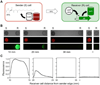
Figure 3
Distance dependence of AHL-mediated cell-cell signaling. (A) Sender E. coli constitutively synthesize the AHL N-3-oxo-dodecanoyl-L-homoserine lactone, which diffuses into the environment and actives GFP expression when receiver cells are sufficiently ...
The varying rates of AHL sorption by low and high temperature biochars suggested that the surface area and adsorption capacities of each biochar, which are determined in part by pyrolysis temperature, would influence the extent to which biochars interfere with microbial communication. To test this idea, we examined how signaling between sender and receiver cells growing on agar plates was altered when the AHL signal produced by sender cells had to diffuse through agar containing the biochars that displayed the largest differences in AHL sorption, 300 and 700°C materials. Since pyrolyzed organic matter strongly absorbs the wavelengths of light used for GFP excitation and emission, we separated sender and receiver cells using a cuboid slab (8 × 37 × 3.4 mm) of the agar medium (1 mL) containing a low concentration (10 mg/mL) of biochar particles (Fig. S6). Biochar occupied only a very small volume fraction (less than 2%) of these agar slabs and should have a negligible effect on the diffusion coefficient of AHL. This arrangement allowed for imaging of GFP expression within receiver cells without biochar interference. To control for signaling variation arising from plate to plate variability in sender cell density, e.g., differences in number of sender cells spotted or variability in the temperature experienced within the incubator, signaling through an agar slab lacking biochars was assayed on the same plate by placing a second agar slab and spot of receiver cells on the opposite side of the sender cells (Fig. 4A). This allowed us to avoid error associated with this variability by never comparing the fluorescence intensities between individual plates, but instead comparing the relative signals from the two spots of receiver cells on each plate.
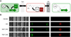
Figure 4
Biochars inhibit cell-cell communication to differing extents. (A) Agar plate assay for assessing biochar effects on E. coli sender-receiver communication. Sender cells were spotted between two agar slabs, one lacking biochar and one containing biochar ...
Visible images show that receiver cells grew to similar extents when proximal and distal from biochars (Fig. 4B), and red fluorescence images reveal constitutive expression of LasI-mCherry within all of the sender cells. These findings show that biochars do not interfere with bacterial growth or alter gene expression that is not regulated by AHL. However, receiver cells grown adjacent to both low and high temperature biochars displayed decreased green fluorescence compared to cells grown next to empty agar. We quantified the effect of each biochar on cell-cell communication by calculating the ratio of GFP expression within the two spots of receiver cells on each plate (Fig. S7). In the case of the low temperature biochar, the receiver cells adjacent to the biochar exhibited 24.1 ±2.1% of the GFP fluorescence observed in the receiver cells grown adjacent to agar lacking biochar. In contrast, the receiver cells grown adjacent to high temperature biochar (700°C) displayed 2.2 ±1.5% of the GFP fluorescence observed within the receiver cells grown adjacent to empty agar. These findings provide the first direct evidence that biochars are capable of altering protein expression that depends on intercellular signaling, and they show that biochar-induced changes in protein expression can vary by an order of magnitude.
Implications for biochar use
We have shown that biochars produced at different temperatures vary in the extent to which they disrupt cell-cell communication among growing bacteria involving one type of AHL. The biochar concentration that elicited this effect (1% by mass) is comparable to the levels that generate biological effects within soils.5,53 This work has implications for carbon sequestration following biochar amendment to soils. Biochars that interfere with microbial conversations could directly alter soil respiration by changing the extent to which microbes form and disperse organic matrices on soil surfaces42,54 and could trigger agronomic effects by altering the fraction of microbes that infect plants and form soft rot.55 Biochar interference with the intercellular signaling needed to trigger root infections may be an economically sought-after agronomic effect, and it may be possible to engineer biochars to intentionally trigger this interference. Alteration of biofilm formation may decrease or increase net CO2 emissions, suggesting additional biochar engineering targets. Biochar amendment could also influence carbon fixation by altering signaling that nitrogen-fixing microbes use to establish and maintain symbiosis with plants,42which is critical in legumes for generating above ground biomass. A similar mechanism could underlie the recent observation that biochar and nutrient conditions exist under which mycorrhizal fungi switch from being mutualists of sorghum to being parasites.25
When preparing biochar, many parameters likely influence its ability to sorb biological molecules, including the feedstock, temperature, minerals, oxygen, and reactor type.56,57 These parameters remain poorly constrained, limiting our ability to predict biochar properties upon amendment to soils. The microbial assay described here will be useful as a simple screen to characterize how these parameters influence biochar sorption of a single biological signaling molecule. Although the details of how biochar sorbs organic compounds are not completely understood, a number of studies indicate that the sorption of organics onto biochar is only partially reversible and can be saturated (as reviewed in Smernik30). This suggests that biochar’s effects on microbial signaling should occur as a short-term pulse associated with the addition of fresh char into soils, and should decline as the char surface is saturated with organic molecules. Evidence exists that biochar particles become oxidized through both abiotic58,59 and biotic25 processes. Biochars also change adsorption capacity with simulated aging. Biochars can decrease in their capacity to absorb nonpolar molecules when they become loaded with lipids that block the access to interior pore networks,60 and they can increase in ion exchange capacity.61 Even if the observed sorption trends are transient within soils, short-lived biochar amendment effects within an ecosystem may be sufficient to drive changes in the trajectory of microbial community development, particularly if biochar is added at the time of seeding or seeding establishment. Charcoal from fires may have similar effects, as indicated by observations of short term microbial impacts from natural charcoal on soil microbial processes.23,62 Further analysis will be needed to determine if the biochar trends observed here are altered by weathering in the environment.
The experimental strategy described here illustrates how synthetic biology63 can aid in constructing microbial assays to characterize the biological processes that drive complex ecological effects from biochar and other soil materials. Microbial biosensors will be useful for determining whether the trends we observe apply to the natural diversity of AHL used for quorum sensing, which have acyl chains of varying lengths,43 as well as other classes of diffusible signaling molecules, such as those used for intraspecies34,35 and interspecies36,38 communication in bacteria (e.g., furanosyl borate diesters, indole, oligopeptides, and quinolones) and fungi (e.g., farnesol, tyrosol, and dimethoxycinnamate). For a given biochar, signaling molecule sorption is expected to vary from organism to organism because of differences in the structures of the molecules used for intercellular communication. Organisms using multiple signaling molecules for cell-cell communication may have only a subset of their intercellular conversations disrupted by a biochar. While our work focused on the effects of biochar on these intercellular conversations, the methods described here could also be used to test the single and combined effects of a wide range of soil materials on microbial communication, and our biochar results will ultimately benefit from being placed in this broad context.
A better understanding of biochar interactions with individual biochemical signals will help constrain parameter space in greenhouse experiments that seek to dissect the complex biotic effects caused by biochar and require significant investments in time and space to achieve replication. Such information will be critical for anticipating the biological effects of soil biochar amendment and making land use decisions that maximize agricultural productivity and carbon sequestration while minimizing greenhouse gas emissions.
Charcoal has a long soil residence time, which has resulted in its production and use as a carbon sequestration technique (biochar). A range of biological effects can be triggered by soil biochar that can positively and negatively influence carbon storage, such as changing the decomposition rate of organic matter and altering plant biomass production. Sorption of cellular signals has been hypothesized to underlie some of these effects, but it remains unknown whether the binding of biochemical signals occurs, and if so, on time scales relevant to microbial growth and communication. We examined biochar sorption of N-3-oxo-dodecanoyl-L-homoserine lactone, an acyl-homoserine lactone (AHL) intercellular signaling molecule used by many gram-negative soil microbes to regulate gene expression. We show that wood biochars disrupt communication within a growing multicellular system that is made up of sender cells that synthesize AHL and receiver cells that express green fluorescent protein in response to an AHL signal. However, biochar inhibition of AHL-mediated cell-cell communication varied, with the biochar prepared at 700°C (surface area of 301 m2/g) inhibiting cellular communication 10-fold more than an equivalent mass of biochar prepared at 300°C (surface area of 3 m2/g). These findings provide the first direct evidence that biochars elicit a range of effects on gene expression dependent on intercellular signaling, implicating the method of biochar preparation as a parameter that could be tuned to regulate microbial-dependent soil processes, like nitrogen fixation and pest attack of root crops.
Keywords: biochar, carbon sequestration, charcoal, microbial communication
Go to:
Introduction
Charcoal is a ubiquitous component of the Earth system, present not only in the soils of fire-affected ecosystems, but also in marine sediments and terrestrial and marine dissolved organic carbon pools.1 It also forms the basis for a form of carbon sequestration called soil biochar amendment,2 where charcoal is intentionally added to soil with the aim of increasing the size of the stable organic carbon pool while potentially delivering other agronomic benefits such as increased crop productivity,1,3–5 decreased soil tensile strength,2,6 altered soil water properties,7–10 improved plant pest resistance,11 and decreased nitrogen loss from soils.12 The carbon sequestration potential of biochar is based the assumption that a large fraction of this material is an inert component of the soil organic matter pool with a very long soil residence time.13 Large-scale data on the biogeochemical cycling of charcoal back up the idea that it decomposes slowly in the Earth system,14,15 and laboratory and ecosystem studies are narrowing down the factors that control charcoal soil residence time.16–20
As commercial applications of biochar have begun, results have appeared which challenge the inert nature of this material and suggest more complex carbon cycle roles. For example, biochar addition to soil has been shown to promote the loss of non-charcoal organic matter (priming) in some studies,21 but not in others.22 This priming of soil decomposition causes an enhanced flux of CO2 into the atmosphere from soils, potentially decreasing the carbon sequestration benefits of biochar, as well as decreasing ecosystem services provided by soil organic matter. Other evidence has appeared suggesting that biochar can enhance retention of nitrogen molecules that contribute to soil fertility,23,24 stimulate colonization of roots by mycorrhizal fungi,25,26 change soil microbial composition,27 and confer plant resistance to microbial pathogens.28 These biological effects have been proposed to arise in part because biochars sorb diffusible small molecules that soil organisms use for intercellular communication and coordinated decision-making.25,29Biochars are known to bind diverse organic molecules,30 many of which are nonpolar like the molecules used for intercellular communication. Numerous studies have analyzed the kinetics of biochar sorption to organics on time scales (days to months) relevant to the mobilization of pollutants.29,31–33 However, it remains unclear if communication can be altered by the presence of biochars on the time scales of microbial signaling and gene expression (minutes to hours).
It is challenging to directly demonstrate biochar-driven mechanisms for observed biological effects within the environment because of the complexity and diversity of conversations occurring among microbes and plants. Bacteria communicate with one another using a variety of biochemicals,34 which are distinct from the molecules used by fungi for intraspecies communication.35 Plants also synthesize flavinoids that regulate microbial behaviors, such as the establishment of root nodules,36 and microbes synthesize nodulation signals37 and plant hormones38 that influence plant development and nutrient uptake. The diversity of signals present in the environment creates challenges in attributing causality to any particular intercellular conservation. The high light absorptivity of biochar and soils adds to this challenge. While there exist microbial biosensors capable of synthesizing reporters that can be imaged when they encounter biological signals within the rhizosphere,39,40fluorescent reporters tend to absorb and emit light in a visible range that is unsuitable for imaging cells within biochar-amended materials, which have high light absorption.
To better understand the effects of biochars on cellular communication, we investigated how biochar materials created under different pyrolysis temperatures (300, 350, 400, 450, 550, 600, and 700°C) influence signal detection within a synthetic microbial system.41 Because the relevant question is the detectability of molecular signals by microbes (as opposed to the chemically-extractable fraction of signal present in the soil), we used a microbial sensor to determine when signaling molecules fell below a level detectable to bacteria in the presence of different biochars. We focused our attention on N-3-oxo-dodecanoyl-L-homoserine lactone, a member of the acyl-homoserine lactone (AHL) signaling molecule family that are used for intraspecies communication and quorum sensing by many gram-negative bacteria, including nitrogen fixing plant symbionts and pathogens that cause soft rot in plants.42,43
Go to:
Materials and Methods
Materials
Escherichia coli XL1-Blue were from Stratagene, and E. coli BLIM cells were kindly provided by K.S. Matthews.44 The acylhomoserine lactone (AHL) used, N-3-oxo-dodecanoyl-L-homoserine lactone, was from Cayman Chemical, bacterial growth media components were from BD Biosciences, and all other reagents were from Sigma-Aldrich and VWR.
Biochar synthesis
Slow pyrolysis of Prosopis glandulosa (mesquite) wood to generate biochar was performed using a fixed bed reactor as described previously.7 In brief, mesquite feedstocks ground to 20 mesh (<0.853 mm) were placed in a stainless steel crucible, which was then plugged with ceramic wool, capped with a ceramic bowl, and buried in fine-grained quartz sand inside a larger, open-top stainless steel crucible. This reactor system was heated in a muffle furnace at 5°C min−1 to the desired reaction temperatures (300, 350, 400, 450, 550, 600, and 700°C) and held at each temperature for 4 hours. The products were manually mixed after cooling to increase homogeneity.
Surface area measurements
We measured the biochar surface area using a Quantachrome Autosorb-3b Surface Analyzer. Prior to analysis, samples were placed in ashed (550°C for 4 hours) glass cells and vacuum dried overnight at 200°C. Nitrogen adsorption/desorption isotherms were obtained at 77 K by a 26-point analysis for relative pressures P/P0from 1.21 × 10⁻⁴ to 0.99, where P is the adsorption equilibrium pressure and P0 is the vapor pressure of bulk liquid N2 at the experimental temperature. Specific surface area was calculated using Brunauer-Emmett-Teller (BET) theory.45
Receiver plasmids
Receiver plasmids contained the synthetic PlasR promoter fused to a strong RBS (BBa_B0034) and GFP gene (BBa_E0040) from the IGEM registry. These plasmids additionally contained a pMB1 origin from pET28a and either a chloramphenicol (CmR) or a kanamycin (KanR) selectable marker.
Sender plasmid
The sender plasmid contained the PA1lacO-1 promoter46 fused to a strong RBS and a gene fusion that was made up of the LasI gene (BBa_C0078) fused to the mCherry gene through a (GGGGS)3 peptide linker. This plasmid additionally contained a pMB1 origin, a CmR marker, and the LacI gene from pET28a. In this sender plasmid, the N-3-oxo-dodecanoyl-L-homoserine lactone synthase gene, LasI, is expressed as a fusion to mCherry through a (GGGGS)3 linker using a PA1lacO-1promoter, which is constitutively active in cells that lack the repressor LacI, such asE. coli BLIM cells.44 The function of this fusion protein is indistinguishable from LasI.
Effect of AHL on GFP expression in liquid culture
E. coli XL1 Blue harboring the receiver plasmid (CmR) were grown to stationary phase in LB medium containing 34 Mg/mL chloramphenicol, diluted to an A600 = 0.05 in 50% LB medium that contained varying concentrations of AHL (0, 5, 25, 50, 125, 250, 500, 1000, and 2000 nM), and grown to stationary phase at 30°C. GFP levels were determined by measuring whole cell fluorescence emission (509 nm) upon excitation of 488 nm using a Tecan M1000 plate reader. Fluorescence was normalized to culture absorbance at 600 nm to account for variability in growth.
Effect of biochar on AHL availability in water
Varying concentrations of each biochar (1, 5, 10, 25, and 50 mg/mL) prepared at different temperatures (300, 350, 400, 450, 550, 600, and 700°C) were incubated with 1 MM AHL within water for 6, 60, or 1440 minutes. After incubation, biochar was removed through centrifugation (14,000 g for 1 min), and 100 ML of the soluble fraction was mixed with an equal volume of LB medium containing E. colitransformed with the CmR receiver plasmid at an OD600 = 0.05 and 50 Mg/mL chloramphenicol. This mixture was grown for 18 hours within 96-well microtiter plates incubated at 30°C and shaken at 250 rpm. Green cellular fluorescence (λex = 488; λem = 509) and absorbance (600 nm) were measured using 200 ML of each culture after growing cells in a shaking incubator (30°C; 250 rpm) for 18 hours. To account for variation in cell density, fluorescence to absorbance ratio was calculated for each well, and the calculated values were normalized to those observed with cells that were grown in the absence of AHL and in the presence of AHL that was not incubated with a biochar.
Effect of colony separation on intercellular signaling
E. coli XL1 Blue harboring the KanR receiver plasmid and E. coli BLIM transformed with the CmR sender plasmid were grown to stationary phase in LB containing 50 Mg/mL kanamycin and 50 Mg/mL chloramphenicol, respectively. Bacteria (1 mL) were harvested by centrifugation, washed with 1 mL of 25% glycerol, resuspended in 1 mL of 25% glycerol, and diluted 10-fold in 25% glycerol. Resuspended receiver (8 ML) and sender cells (3 ML) were spotted at different distances (1, 10, 20, and 30 mm) from one another on 1.5% agar plates containing M9 minimal medium and twenty amino acids. A smaller volume of the sender strain was spotted to avoid growth of these cells over the biochar agar slabs during the overnight incubation. Spotting was performed in triplicate at each separation distance. After spotting, plates were incubated overnight at 37°C and then photographed with a Canon Digital Rebel mounted on a Leica MZFLIII microscope at 0.4x magnification with white light (5 ms exposure), a Chroma 41012 GFP long pass filter set (200 and 500 ms exposures), and a Chroma 11002 Green filter (200 and 500 ms exposures). The image analysis software ImageJ was used to quantify the fluorescence from receiver colonies on each agar plate.47
Effect of biochar on cell-cell communication on solid medium
Agar plates for assessing cellular communication were prepared by placing a mold (sterilized through immersion in ethanol) within sterile polystyrene plates (60×15 mm) and pouring 8 mL of 1.5 agar containing M9 medium and twenty amino acids. Upon cooling the mold was removed to leave a pair of empty adjacent wells. The left well (control) on each plate was filled with 1 mL liquid agar (1.5%) dissolved in water (65°C). The right well (experimental) was then filled with 1 mL liquid agar containing or lacking 10 mg of 300°C or 700°C biochar. E. coli XL1 Blue harboring the KanR receiver plasmid and E. coli BLIM transformed with the CmR sender plasmid were grown to stationary phase in LB containing 50 Mg/mL kanamycin and 50 Mg/mL chloramphenicol. Bacteria were harvested by centrifugation, washed with 25% glycerol, resuspended in 25% glycerol, and diluted 10-fold into 25% glycerol. The 10-fold dilution of sender cells (3 ML) was spotted between the two agar slabs on plates, whereas the 10-fold dilution of receiver cells (8 ML) was spotted on the outside of each agar slab. After spotting, plates were incubated overnight at 37°C and then photographed with a Canon Digital Rebel on a Leica MZFLIII microscope at 0.4× magnification with white light (5 ms exposure), a Chroma 41012 GFP long pass filter set (200 and 500 ms exposures), and a Chroma 11002 Green filter (200 and 500 ms exposures). ImageJ was used to quantify the fluorescence from the pair of receiver colonies on each agar plate. The ratio of the signals from the pair of receiver colonies on each plate was calculated (receiver adjacent to biochar/receiver adjacent to empty agar) to determine the level of signal inhibition elicited by each biochar. All measurements were performed in triplicate, and values reported represent the average of three independent experiments ±1σ.
Calculating the effect of biochar on AHL diffusion
To estimate the effect of biochar on AHL diffusion within agar plates, the effective diffusion coefficient De of AHL was calculated as (1 − ε)D0/[1 − 0.5 ln(1 − ε)], where D0 is the actual diffusion coefficient in biochar-free agar medium and is the volume fraction occupied by biochar particles in the agar-BC cube.48 This analysis revealed that the low volume fraction used in our experiments (less than 2% v/v biochar) decreases the AHL diffusion constant by less than 3%. This implicates sorption as the major mechanism responsible for biochar effects on sender-receiver cell-cell signaling.
Go to:
Results and Discussion
Autoinducer sorption to biochars
To assay whether biochars influence the availability of AHLs, we constructed a bacterial biosensor39 that expresses a green fluorescent protein (GFP) reporter when bacterial cells encounter the AHL N-3-oxo-dodecanoyl-L-homoserine lactone in their local environment. This was accomplished by transforming E. coli with a plasmid that: i) constitutively expresses the protein LasR, a Pseudomonas aeruginosa AHL-dependent protein that activates transcription from the promoterPlasR, and ii) expresses GFP under control of the LasR-dependent promoter PlasR. Cells containing this receiver plasmid displayed increased GFP production when they were grown in medium containing AHL (Fig. S1), because LasR requires bound AHL to activate transcription of genes whose expression is regulated by the LasR promoter.49 We assayed AHL signaling in E. coli because this organism does not use AHL to communicate, lacks the complex responses of soil bacteria that are sensitive to one or more AHL, and does not produce lactonase enzymes that hydrolyze AHL. With this bacterial biosensor, only two parameters determine AHL levels in the medium: the amount of AHL added to the medium and fraction of the AHL that sorbs to biochar.
We produced a suite of biochars by pyrolyzing Prosopis glandulosa (mesquite) wood chips over a range of temperatures (300, 350, 400, 450, 550, 600, and 700°C). N2 sorption analysis of these biochars revealed surface areas that spanned more than two orders of magnitude (Fig. S2). Varying amounts of each biochar (1 to 50 mg/mL) were incubated with a concentration of AHL (1 MM) sufficient to activate E. coli to synthesize GFP (Fig. S1). This level of AHL is within a range that is thought to be biologically relevant for Pseudomonas aeruginosa.50 Initial incubations were performed on the timescale of E. coli reproduction. After 60 minutes, the biochar was separated from unbound AHL by centrifugation, the soluble fraction was mixed with a low density culture of E. coli, and cells were grown to stationary phase to allow for GFP expression. Whole cell GFP fluorescence measurements revealed that incubation of AHL with the highest concentrations of each biochar decreased AHL-dependent cellular fluorescence in all cases (Fig. 1). The concentration of biochar required to suppress cellular fluorescence varied with biochar pyrolysis temperature and surface area. The biochars with similar low surface areas (300, 350, and 400°C) required the highest concentrations to suppress GFP expression half maximally (10–50 mg/mL), the biochars with the highest surface areas (550, 600, and 700°C) required the lowest concentrations to suppress GFP expression half maximally (1–2 mg/mL), and the biochar with an intermediate surface area (450°C) required an intermediate level of biochar (5 mg/mL) to suppress GFP expression half maximally. We also found that the correlation between biochar surface area and the concentration required for half maximal inhibition of GFP fluorescence could be described by a simple exponential model, R2 = 0.99 (Fig. 2). This trend implicates the AHL binding capacity of the biochar as dependent on surface area and pyrolysis temperature under the conditions used to produce our biochars. Cells incubated with biochar-treated AHL all grew to a similar maximal density (Fig. S3), indicating that biochar-treated water did not alter cell growth.
Figure 1
Sorption of AHL by biochars pyrolyzed over a range of temperatures. (A) E. coli transformed with the receiver plasmid synthesize GFP when grown in an environment containing the AHL N-3-oxo-dodecanoyl-L-homoserine lactone. The transcriptional activator ...
Figure 2
Relationship between biochar surface area, production temperature, and inhibition of GFP reporter expression. The concentration of biochar required to decrease AHL-induced GFP fluorescence by 50% within whole cells was estimated from the data presented ...
We also sought to establish whether the biochar sorption of AHL had reached equilibrium with our low surface area biochars and whether biochar sorption occurred on an even faster time scale with our high surface area biochars, e.g., faster than the doubling time of E. coli in rich growth medium. If some biochars sorb AHL on the timescale of very rapidly-growing E. coli under optimal growth conditions in a lab, they are likely to sorb AHL under the lower nutrient, slower growth conditions experienced by soil microbes in the environment. To address these questions, we varied the incubation time of AHL and biochar prior to mixing the soluble fraction with E. coli. Incubation of low temperature biochars (300 to 400°C) with AHL for 24 hours showed a greater inhibition of GFP expression than 1 hour incubation (Fig. S4), indicating that these reactions had not reached equilibrium after one hour. The lack of saturation suggests the ability of biochar to cause a continued suppression of microbial signaling as bacterial populations grow. In addition, AHL-induced cellular fluorescence was suppressed when the high temperature biochars (550 to 700°C) were incubated with AHL for only six minutes prior to mixing with E. coli (Fig. S5). This latter finding suggested that some biochars are capable of adsorbing AHL on the very shortest time scales of microbial reproduction and signaling within rich growth medium, a condition that likely represents the upper bound on the rate of growth and signaling within the environment.
Effect of biochars on AHL-mediated cell-cell communication
To directly assess whether biochars can disrupt communication among growing bacteria, we built a sender plasmid that programs E. coli to express P. aeruginosaLasI (Fig. 3A), the acyl-homoserinelactone synthase that continuously synthesizes the AHL N-3-oxododecanoyl-L-homoserine lactone from bacterial metabolites.51 LasI was expressed as a fusion to the red fluorescent protein mCherry to allow for visualization of expression. To make LasI production of N-3-oxo-dodecanoyl-L-homoserine lactone continuous within sender cells, we used a PA1lacO-1 promoter to constitutively express the LasI-mCherry fusion protein and E. coli BLIM cells that are devoid of the transcriptional lac repressor, which inhibits expression of genes from this promoter. To determine how receiver-sender cell-cell communication varied with distance within a synthetic multicellular system,41 E. coli cells harboring each plasmid type were spotted onto solid growth medium at different edge-to-edge distances from one another (10 to 30 mm), and cell density and GFP expression levels were imaged after overnight growth using visible and fluorescence microscopy, respectively (Fig. 3B). Cells harboring each plasmid displayed similar growth under all conditions analyzed. However, the cells containing the receiver plasmid displayed GFP fluorescence that was inversely related to the distance between the sender and receiver cells. When sender and receiver cells were separated by 10 to 20 mm, a gradient of GFP expression was observed within the receiver cells (Fig. 3C). While this separation is greater than the distance where signaling is thought to occur on plant surfaces in natural systems,52 it was necessary for imaging the GFP reporter signal without biochar interference.
Figure 3
Distance dependence of AHL-mediated cell-cell signaling. (A) Sender E. coli constitutively synthesize the AHL N-3-oxo-dodecanoyl-L-homoserine lactone, which diffuses into the environment and actives GFP expression when receiver cells are sufficiently ...
The varying rates of AHL sorption by low and high temperature biochars suggested that the surface area and adsorption capacities of each biochar, which are determined in part by pyrolysis temperature, would influence the extent to which biochars interfere with microbial communication. To test this idea, we examined how signaling between sender and receiver cells growing on agar plates was altered when the AHL signal produced by sender cells had to diffuse through agar containing the biochars that displayed the largest differences in AHL sorption, 300 and 700°C materials. Since pyrolyzed organic matter strongly absorbs the wavelengths of light used for GFP excitation and emission, we separated sender and receiver cells using a cuboid slab (8 × 37 × 3.4 mm) of the agar medium (1 mL) containing a low concentration (10 mg/mL) of biochar particles (Fig. S6). Biochar occupied only a very small volume fraction (less than 2%) of these agar slabs and should have a negligible effect on the diffusion coefficient of AHL. This arrangement allowed for imaging of GFP expression within receiver cells without biochar interference. To control for signaling variation arising from plate to plate variability in sender cell density, e.g., differences in number of sender cells spotted or variability in the temperature experienced within the incubator, signaling through an agar slab lacking biochars was assayed on the same plate by placing a second agar slab and spot of receiver cells on the opposite side of the sender cells (Fig. 4A). This allowed us to avoid error associated with this variability by never comparing the fluorescence intensities between individual plates, but instead comparing the relative signals from the two spots of receiver cells on each plate.
Figure 4
Biochars inhibit cell-cell communication to differing extents. (A) Agar plate assay for assessing biochar effects on E. coli sender-receiver communication. Sender cells were spotted between two agar slabs, one lacking biochar and one containing biochar ...
Visible images show that receiver cells grew to similar extents when proximal and distal from biochars (Fig. 4B), and red fluorescence images reveal constitutive expression of LasI-mCherry within all of the sender cells. These findings show that biochars do not interfere with bacterial growth or alter gene expression that is not regulated by AHL. However, receiver cells grown adjacent to both low and high temperature biochars displayed decreased green fluorescence compared to cells grown next to empty agar. We quantified the effect of each biochar on cell-cell communication by calculating the ratio of GFP expression within the two spots of receiver cells on each plate (Fig. S7). In the case of the low temperature biochar, the receiver cells adjacent to the biochar exhibited 24.1 ±2.1% of the GFP fluorescence observed in the receiver cells grown adjacent to agar lacking biochar. In contrast, the receiver cells grown adjacent to high temperature biochar (700°C) displayed 2.2 ±1.5% of the GFP fluorescence observed within the receiver cells grown adjacent to empty agar. These findings provide the first direct evidence that biochars are capable of altering protein expression that depends on intercellular signaling, and they show that biochar-induced changes in protein expression can vary by an order of magnitude.
Implications for biochar use
We have shown that biochars produced at different temperatures vary in the extent to which they disrupt cell-cell communication among growing bacteria involving one type of AHL. The biochar concentration that elicited this effect (1% by mass) is comparable to the levels that generate biological effects within soils.5,53 This work has implications for carbon sequestration following biochar amendment to soils. Biochars that interfere with microbial conversations could directly alter soil respiration by changing the extent to which microbes form and disperse organic matrices on soil surfaces42,54 and could trigger agronomic effects by altering the fraction of microbes that infect plants and form soft rot.55 Biochar interference with the intercellular signaling needed to trigger root infections may be an economically sought-after agronomic effect, and it may be possible to engineer biochars to intentionally trigger this interference. Alteration of biofilm formation may decrease or increase net CO2 emissions, suggesting additional biochar engineering targets. Biochar amendment could also influence carbon fixation by altering signaling that nitrogen-fixing microbes use to establish and maintain symbiosis with plants,42which is critical in legumes for generating above ground biomass. A similar mechanism could underlie the recent observation that biochar and nutrient conditions exist under which mycorrhizal fungi switch from being mutualists of sorghum to being parasites.25
When preparing biochar, many parameters likely influence its ability to sorb biological molecules, including the feedstock, temperature, minerals, oxygen, and reactor type.56,57 These parameters remain poorly constrained, limiting our ability to predict biochar properties upon amendment to soils. The microbial assay described here will be useful as a simple screen to characterize how these parameters influence biochar sorption of a single biological signaling molecule. Although the details of how biochar sorbs organic compounds are not completely understood, a number of studies indicate that the sorption of organics onto biochar is only partially reversible and can be saturated (as reviewed in Smernik30). This suggests that biochar’s effects on microbial signaling should occur as a short-term pulse associated with the addition of fresh char into soils, and should decline as the char surface is saturated with organic molecules. Evidence exists that biochar particles become oxidized through both abiotic58,59 and biotic25 processes. Biochars also change adsorption capacity with simulated aging. Biochars can decrease in their capacity to absorb nonpolar molecules when they become loaded with lipids that block the access to interior pore networks,60 and they can increase in ion exchange capacity.61 Even if the observed sorption trends are transient within soils, short-lived biochar amendment effects within an ecosystem may be sufficient to drive changes in the trajectory of microbial community development, particularly if biochar is added at the time of seeding or seeding establishment. Charcoal from fires may have similar effects, as indicated by observations of short term microbial impacts from natural charcoal on soil microbial processes.23,62 Further analysis will be needed to determine if the biochar trends observed here are altered by weathering in the environment.
The experimental strategy described here illustrates how synthetic biology63 can aid in constructing microbial assays to characterize the biological processes that drive complex ecological effects from biochar and other soil materials. Microbial biosensors will be useful for determining whether the trends we observe apply to the natural diversity of AHL used for quorum sensing, which have acyl chains of varying lengths,43 as well as other classes of diffusible signaling molecules, such as those used for intraspecies34,35 and interspecies36,38 communication in bacteria (e.g., furanosyl borate diesters, indole, oligopeptides, and quinolones) and fungi (e.g., farnesol, tyrosol, and dimethoxycinnamate). For a given biochar, signaling molecule sorption is expected to vary from organism to organism because of differences in the structures of the molecules used for intercellular communication. Organisms using multiple signaling molecules for cell-cell communication may have only a subset of their intercellular conversations disrupted by a biochar. While our work focused on the effects of biochar on these intercellular conversations, the methods described here could also be used to test the single and combined effects of a wide range of soil materials on microbial communication, and our biochar results will ultimately benefit from being placed in this broad context.
A better understanding of biochar interactions with individual biochemical signals will help constrain parameter space in greenhouse experiments that seek to dissect the complex biotic effects caused by biochar and require significant investments in time and space to achieve replication. Such information will be critical for anticipating the biological effects of soil biochar amendment and making land use decisions that maximize agricultural productivity and carbon sequestration while minimizing greenhouse gas emissions.